AI-powered
Perfusion Analysis

MRAY VEO CORE FÜR FUNDIERTE THERAPIEENTSCHEIDUNGEN

AI Image Analysis: mRay VEO Core

Optimized CT/MRI perfusion analysis

Stroke is one of the leading causes of death and a major cause of severe, permanent disability. Thrombectomy offers a new and improved treatment option. mRay VEO Core supports you in decision-making with automated AI analysis—because “time is brain”!

mRay VEO Core

  • Guideline-compliant fully automated evaluation of perfusion measurements within three minutes.
  • Display DEFUSE-3 inclusion criteria as suggested in ESO Guideline [01.2019]:
    infarct core volume, penumbra volume, mismatch ratio.
  • Intuitive representation in a 3D overview: infarct core (red) and hypoperfusion (yellow) as colored overlay, representation of CBF and Tmax.
  • Automated e-ASPECTS analysis
  • Quantitative results for the quality of the automatic evaluation: warning in case of detected patient movement or incorrect contrast medium injection.
  •  
ki basierte perfusionsanlyse screenshot mray veocore
Full 3D data set with hypoperfusion (TMAX>6s)
ki basierte perfusionsanalyse mRay VEOCORE Motion Correction
Motion correction: warning when patient movement is detected

Fast communication

Thanks to full integration in mRay, all parties involved are informed in real time and can coordinate on the case regardless of location, of course also via mobile devices. Therapy decisions are made quickly and still evidence-based according to the European Stroke Organisation (ESO).

The acquisition is cost-effective, available as a standalone or as a package.

Learn more about mRay VEO Core

mRay VEO Core on deepcOS®

mRay VEO Core is now available via the AI platform deepcOS® and the AI Marketplace – centrally integrated into a growing ecosystem of validated AI solutions for hospitals and radiology facilities.

Learn more

Development with users

mRay VEO Core is a joint project by VEObrain, the University Hospital Freiburg, and mbits—developed on-site according to user needs. Integration into mRay and rapid image data exchange enable optimal stroke care. Because: Time is Brain!

Latest updates on mRay VEO Core

New webinar available: Dr. Ingmar Gergel shares exciting insights into the latest mRay VEO Core features.

  • Perfusion Analysis with mRay VEO Core
  • Outputs and Demo with mRay VEO Core
  • Analysis of Results
  • e-ASPECTS on NCCT
  • Multimodal Stroke Imaging
  • Radiation Reduction

Automated analyses: everything at a glance with mRay

CBF/Mismatch mit MT Indication mRay VEOcore
mRay VEO Core: CBF/Mismatch mit MT Indikation
Quality control: bolus detection and patient movement

Recommended by Experts

This is what mRay users say

Contact us

Interested in mRay?

Get in touch with us via email or phone – we’re happy to assist you. Alternatively, you can explore mRay yourself and discover the countless ways our medical app can be used in everyday clinical practice.

FAQs about Perfusion Analysis

You may already find some answers here

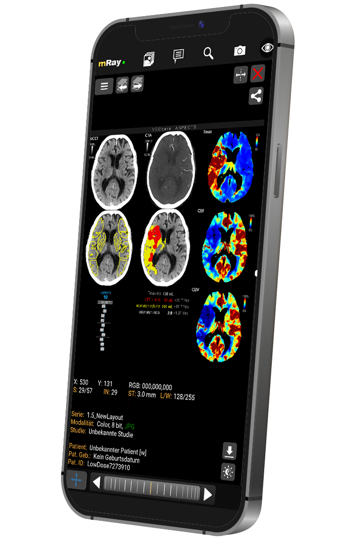
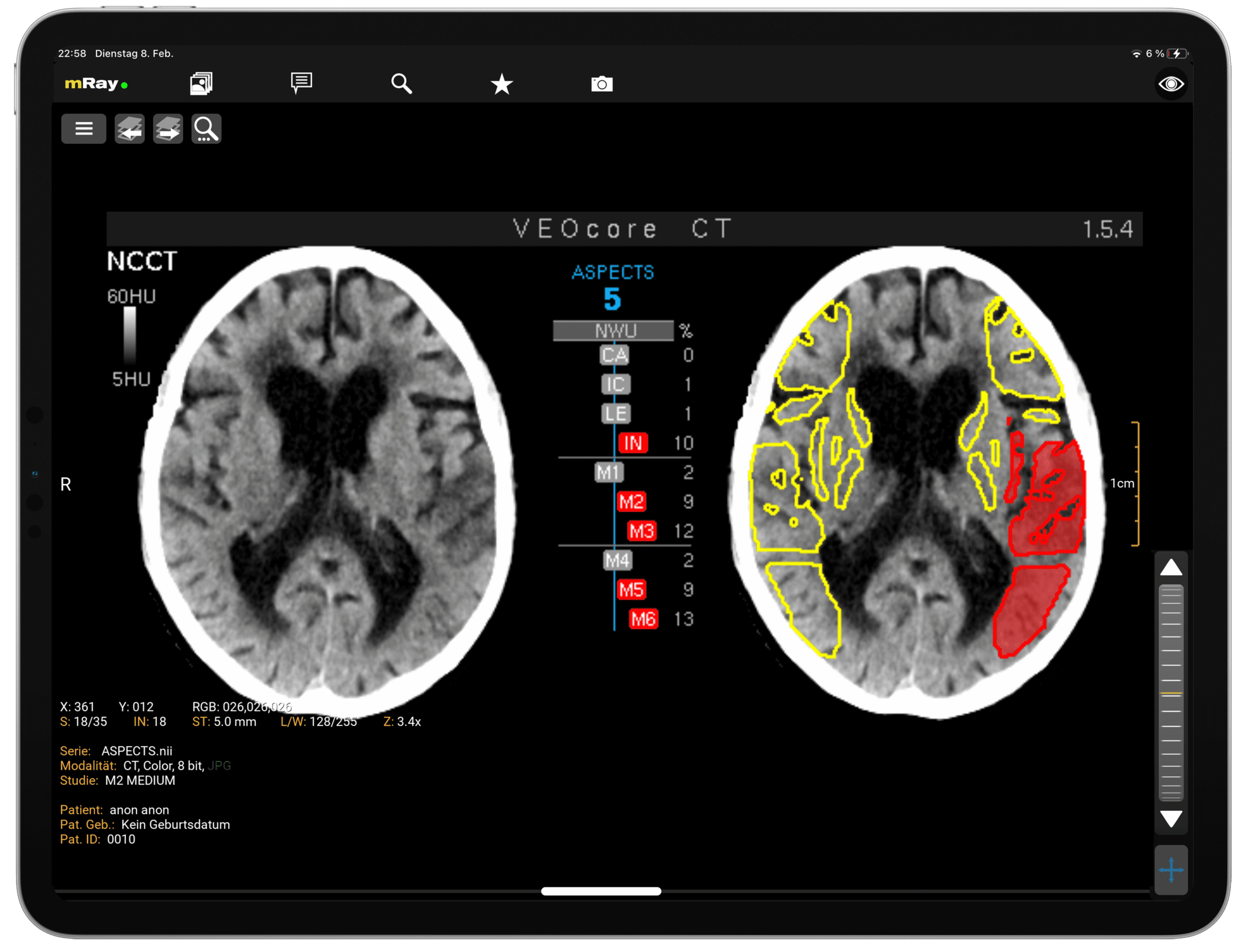
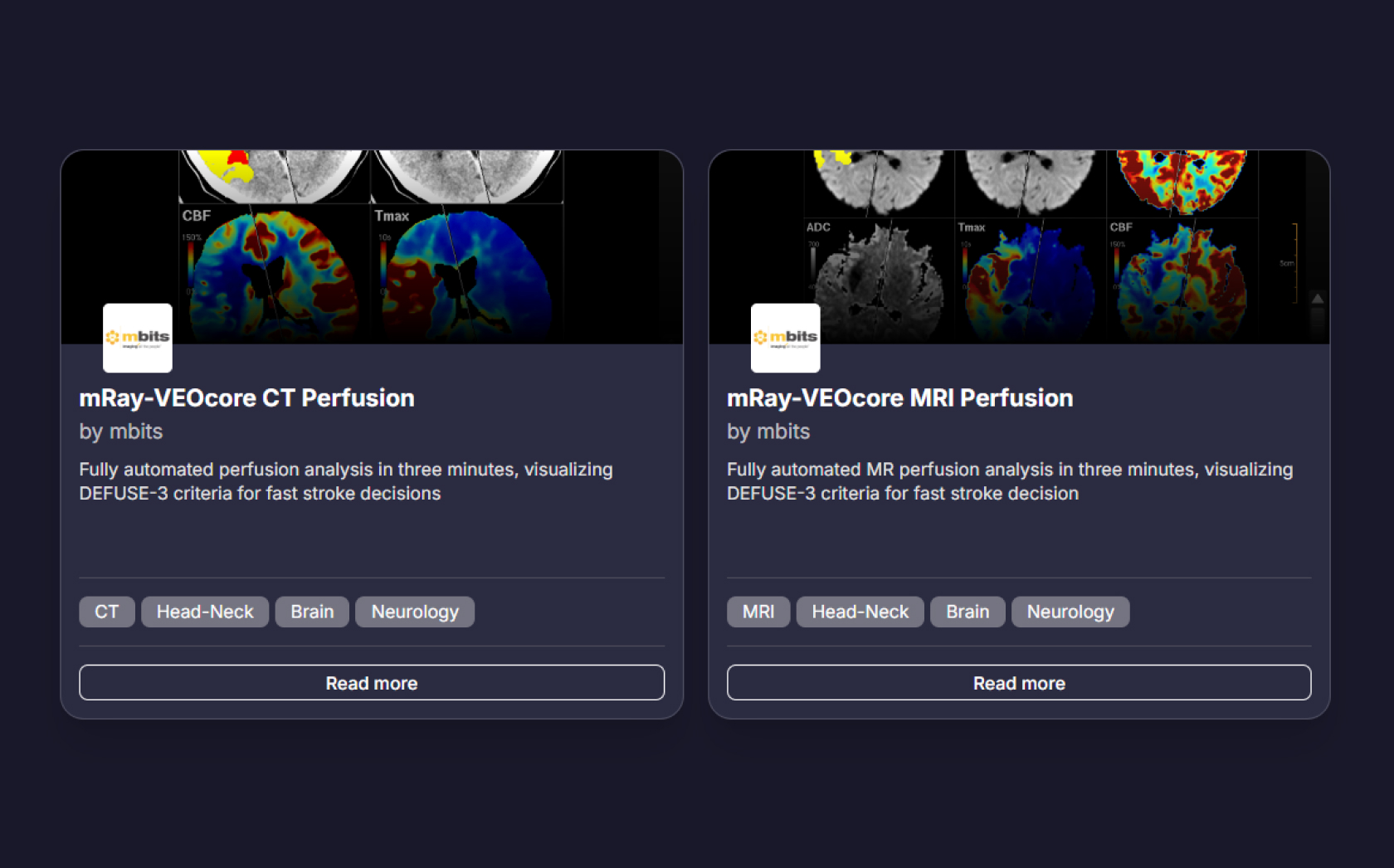
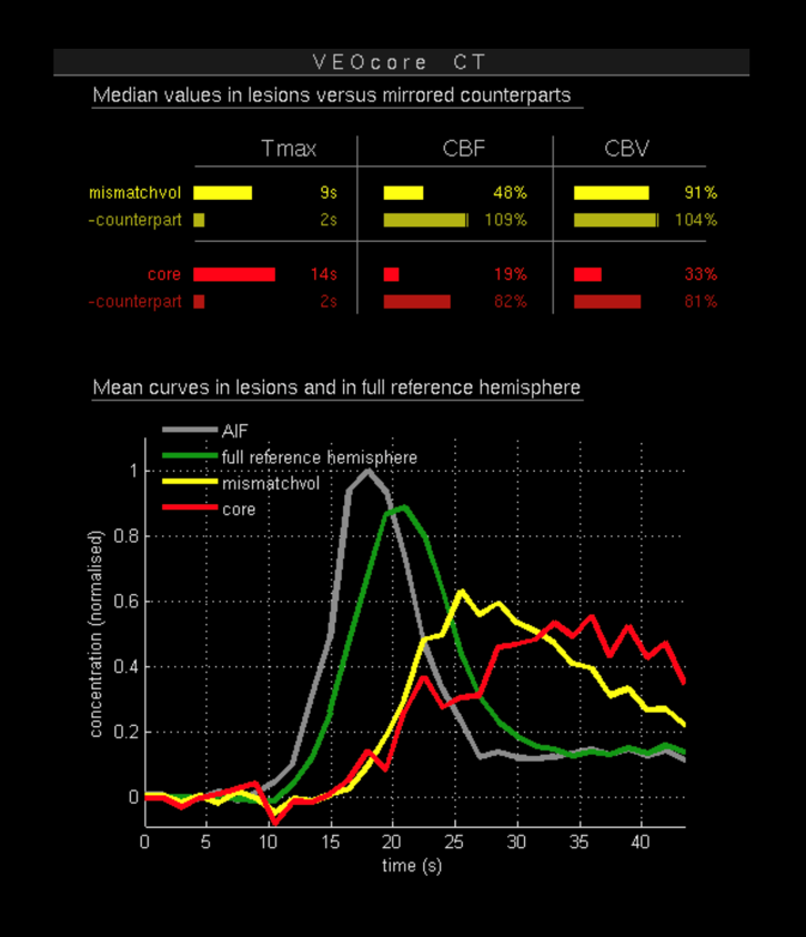
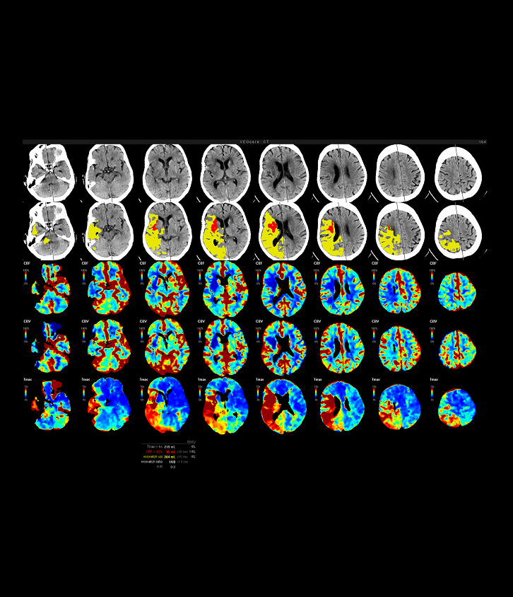
Perfusion analysis of brain images allows visualization and quantification of low-perfusion tissue (penumbra), non-perfusion tissue (core tissue), and the mismatch ratio between the two values. The calculated values can be used to support decision making based on the assessment of the extent of tissue damage.

Usually, the evaluation can be started automatically in the standard configuration. If required, the possibility for manual evaluation can be set up.

EN

Vielen Dank für Ihre Anmeldung

In Kürze erhalten Sie eine E-Mail, mit der Sie das Abonnement unseres Newsletters bestätigen können.