FAQs
HIER FINDEN SIE HILFESTELLUNGEN FÜR MRAY
Übersicht
Stand: 03.08. 2023
mRay FAQ - Anwender*Innen
Zweitmeinung / Betrachtung
JPG bedeutet, dass dieser Datensatz komprimiert (entsprechend der Konsensius-Konferenz) übertragen wird. Siehe auch Loose et al.: Compression of digital images in radiology – results of a consensus conference. Rofo. 2009 Jan;181(1):32-7. doi: 10.1055/s-2008-1027847.
Stellen Sie sicher, dass das Suchfeld leer ist. Durch Löschen der Filtereingabe werden wieder alle Datensätze angezeigt.
Manche PACS Hersteller fordern eine Suchanfrage die Groß- und Kleinbuchstaben unterscheidet. Bsp.: „schmidt“ liefert keine Ergebnisse, „Schmidt“ hingegen schon.
Die Referenzlinien (Schnittebenen) werden anhand der Parameter Image Orientation Patient (0020,0037) und Image Position Patient (0020,0032) aus den DICOM Dateien berechnet. Die Linien werden nur angezeigt, wenn diese Daten vorhanden sind oder sich daraus valide Schnitte berechnen lassen.
mRay bietet die Möglichkeit einen temporären Link auf Studien oder Serien zu erstellen. Dieser kann dem/der Expert*In z.B. per Mail geschickt werden. Der Link ist mit einem PIN vor unerlaubtem Zugriff geschützt.
Ihr Chat Partner hat die Nachricht gelesen, wenn an der Nachricht ein kleines Auge-Symbol gezeigt wird. Die Gelesen-Funktion steht momentan noch nicht in Gruppen zur Verfügung.
- Der angegebene Nutzer existiert nicht.
- Das angegebene Passwort ist nicht korrekt
- Die Groß-Kleinschreibung des Nutzers ist nicht korrekt (Nur bei Versionen vor mRay 6.0)
- mRay in der Klinik: wenden Sie sich an einen IT Mitarbeitenden der Klinik. Er kann Ihnen das Passwort zurücksetzen.
- mRay Cloud: Gehen Sie im Browser auf https://mray.app und wählen Sie im Anmeldebildschirm die Option “Passwort vergessen”. Geben Sie Ihre Email Adresse an, Sie erhalten daraufhin eine Mail mit einem Link zum Zurücksetzen Ihres Passworts.
- mRay in Klinik: Das ist die Adresse (URL) des mRay Servers Ihrer Klinik. Sie erhalten die Adresse von der IT Abteilung Ihrer Klinik.
- mRay Cloud: Verwenden Sie “mray.app”, um mRay Cloud zu verwenden
- mRay in Klinik: Sie erhalten die Adresse von der IT Abteilung Ihrer Klinik
- mRay Cloud: Verwenden Sie “mray.app”, um mRay Cloud zu verwenden
PINs für den Klinik Login werden per Mail an Ihre Klinik Mailadresse verschickt. Stellen Sie sicher, dass Zugriff auf Ihr Postfach haben
- Schauen Sie im Spam Ordner nach. Evtl. wurde die Mail fälschlicherweise als Spam erkannt
- Wenn Sie die Mail nicht in Ihrem Postfach finden können, sprechen Sie Ihre Klinik IT an. Ein*e Mitarbeiter*In kann Ihnen die PIN auch telefonisch / persönlich mitteilen
Ihre Klinik erlaubt aus Sicherheitsgründen den Login nur mit zuvor freigegebenen Geräten. Sprechen Sie eine*n Mitarbeiter*In der Klinik IT an, um das gewünschte Gerät freischalten zu lassen.
Die Ursachen können sehr vielfältig sein. Sie können die aktuelle Übertragungsrate während eines Download im Seitenmenü einsehen, wenn Sie auf das mRay Logo klicken. Prüfen Sie über welche Verbindung Sie gerade verbunden sind, also WLAN oder Mobile Daten.
- Prüfen Sie Ihre Verbindungsart. Sie sind evtl. im Mobilfunknetz und Ihr Datenvolumen ist aufgebraucht.
- Laden Sie evtl. gerade noch andere Sachen außerhalb von mRay herunter? Das kann die Übertragungsrate verringern.
mRay Cloud
- Es kann vorübergehend zu Schwankungen der Datenraten kommen. Sollte das Problem anhalten, wenden Sie sich an support@mbits.info
mRay in der Klinik
- Sollten Sie sich im Klinik WLAN befinden, sprechen Sie Ihre Klinik IT an.
- Führen Sie ggf. einen unabhängigen Geschwindigkeitstest durch. Sollten die erreichten Werte von den Übertragungsgeschwindigkeiten in mRay stark abweichen, sprechen Sie Ihre Klinik IT an.
Teleradiologie
Ja. Mehr dazu finden Sie unter https://mbits.info/wp-content/uploads/2025/04/mRay-Nutzungsanweisungen_Befundung.pdf.
Ja. Dies ist auch ohne mRay interne Kalibrierung möglich.
Wir nehmen Bezug auf folgenden Konsensus und deren Ergebnisse (JPEG-Kompressionsfaktoren). Zitat: “Im Rahmen einer Konsensuskonferenz (Loose et al. 2009) wurde mit Radiologen, Medizinphysikern, Industrie- und Behördenvertretern das Thema Bildkompression von DICOM-Daten behandelt. Auf der Basis der 56 am höchsten bewerteten Studien, die aus 216 Publikationen ausgewählt wurden, sowie der größten aktuell erschienenen Studie aus Kanada diskutierte man, ob und mit welchen Faktoren eine Bildkompression ohne Einschränkung der diagnostischen Bildqualität möglich ist. Das Ergebnis dieser Konferenz, an der mehr als 80 Experten teilnahmen, wurde in der Fachzeitschrift „RöFo – Fortschritte auf dem Gebiet der Röntgenstrahlen und bildgebenden Verfahren“ zu Beginn des Jahres 2009 veröffentlicht.
Die Lux Meter Anzeige ist nur unter iOS verfügbar.
Wenn Sie das Lux Meter geöffnet haben, klicken Sie auf Start, um die Messung durchzuführen. Das Lux Meter bestimmt dann auch die Raumklasse, in der Sie sich befinden und protokolliert diese automatisch.
Die Lux Meter Anzeige ist nur unter iOS verfügbar.
Diese Funktion muss am mRay Server konfiguriert werden und wurde sicher noch nicht eingerichtet. Sprechen Sie Ihre Klinik IT an, sie wird sich für die Einrichtung mit uns in Verbindung setzen.
Diese Funktion muss am mRay Server konfiguriert werden. Sprechen Sie Ihre Klinik IT an, sie wird sich für die Einrichtung mit uns in Verbindung setzen.
Die manuelle Messung der Teil-Strecke zählt zu den Prüfpunkten der Konstanzprüfung, die monatlich erfolgen muss. Eine regelmäßige Messung, bspw. arbeitstäglich, ist empfehlenswert.
Ja, beim Wechseln des Standorts müssen folgende Punkte erneut überprüft werden: initiales Messen der Übertragungszeit (6.2.2), Vollständigkeit der Datenübertragung (6.2.3) (DIN 6868-159).
Fotodokumentation
Nein. Der Zugriff auf Fotos innerhalb der Galerie wird aus Datenschutz- und Sicherheitsgründen nicht unterstützt.
Nein. Die Daten bleiben verschlüsselt innerhalb von mRay und sind nicht durch den Benutzer oder von anderen Apps einsehbar.
VEOcore Perfusionsanalyse
Überprüfen Sie, ob die Hostanwendung oder das Sendegerät (PACS- oder CT/MR-Gerät) eine Warnung oder einen Fehler ausgegeben hat. Wenn nicht, überprüfen Sie sorgfältig, ob Sie alle erforderlichen und die richtigen Eingabebilder gesendet haben. Versuchen Sie, die Bilder erneut zu senden.
Überprüfen Sie den Bewegungskorrekturbericht, um den Schweregrad der Bewegung abzuschätzen und auf Fehlalarme in der Segmentierung zu prüfen. Wenn Sie Zweifel an der Zuverlässigkeit der Ergebnisse haben, sollten Sie diese nicht für Ihre Entscheidungen verwenden.
Überprüfen Sie den Bericht qcontrol_bolus und überprüfen Sie die Zeitkurven. Wenn sie sehr verrauscht sind, interpretieren Sie die Ergebnisse sorgfältig. Wenn in den Zeitkurven kein Bolus sichtbar ist, kann dies auf ein Problem bei der Kontrastmittelinjektion zurückzuführen sein (nicht eingeflossen oder zu spät eingeflossen). In diesem Fall können Sie die Messung wiederholen.
Die Kernsegmentierung basiert auf einem globalen Schwellenwert von 30%. Dies berücksichtigt nicht die Unterschiede in der Grauen und Weißen Substanz. Überprüfen Sie dies visuell mit dem kontralateralen Gegenstück oder im Bericht zu qcontrol_flowstats. Berücksichtigen Sie, dass eine schwere Mikroangiopathie zu einer relevanten Hypoperfusion führen kann, die möglicherweise zu einer falsch positiven CBF-Kernsegmentierung führt. Wenn Sie Zweifel an der Zuverlässigkeit der Ergebnisse haben, sollten Sie diese nicht für Ihre Entscheidungen verwenden.
Sehr kleine Läsionen im Bereich von 1-2mL können durch die Maskengenerierung weggeglättet werden.
In diesem Falle war die Koregistrierung zwischen Perfusion und Diffusion fehlerhaft und Diffusion und Flair wurden fälschlicherweise zur Perfusion verdreht.
Koregistrierungsfehler können leider algorithmisch bedingt passieren (Stichwort “lokales Minimum”).
Grundsätzlich greift bei fehlerbehafteten Auswertungen die im Benutzerhandbuch beschriebene “Fallback”-Strategie:
Visueller Check der Koregistrierung => falls fehlerhaft, visuelle Interpretation der Ergebnisse => falls unklar oder nicht möglich, Auswertung nicht verwenden.
- Es waren gerade noch andere Auswertung im Gange bzw. standen aus. Die Perfusionanalyse mittels VEOcore ist sehr rechenintensiv, deshalb kann mRay meist nur eine Auswertung gleichzeitig durchführen. Bei mehren Auswertungsanfragen kann es deshalb zu Verzögerungen kommen.
- Der Versand der Studie vom PACS war so langsam, dass die Perfusionsauswertung zwischenzeitlich schon gestartet wurde. Leider ist es beim Versand nicht ersichtlich, wann alle Daten angekommen sind. Wird die Logik-Bedingung zum Starten der Auswertung schon vorher erfüllt, kann es unter Umständen zu dieser Problematik kommen. Die fehlerhaft gestartete Auswertung wird dann abgebrochen und neu gestartet. Am Ende sollten deshalb dann aber die richtigen und vollständigen Ergebnisse vorliegen.
– vergleichsweise wenig KM gegeben
– spezielle kardiovaskuläre Situation oder sehr schwerer Patient, so dass wenig KM im Hirn ankommt
– Probleme bei der Injektion (nicht richtig oder alles eingeflossen)
Allgemein
Im Login Feld geben Sie Ihre E-Mail Adresse ein, die Sie bei der Registrierung verwendet haben.
Diese Funktion ist momentan noch nicht vorgesehen. Dies wird in einer zukünftigen mRay Version über In-App Kauf möglich sein.
(Intern: der Speicherplatz kann pro Account / Gruppe über eine Option beliebig gesetzt werden)
Sie müssen der App die Berechtigung erteilen auf das lokale Netzwerk zuzugreifen.
Ausgeblendete Studien und Serien können Sie in den Einstellungen wieder herstellen. Klicken Sie dazu auf den mRay Button oben links und wählen Sie die Einstellungen aus. Sie finden die Option dann unter Geräte Einstellungen >> Daten >> Ausgeblendete Daten anzeigen.
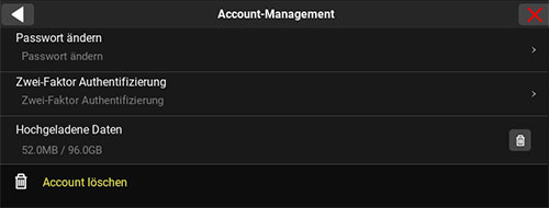
Gehen Sie in der Inbox auf das mRay-Symbol und dann auf Einstellungen. Anschließend rufen Sie das Account-Management auf, dort können Sie Ihr Passwort ändern.
mRay FAQ - ITler*Innen
Nein. Das Gateway ist nur ein Verbindungsvermittler, vergleichbar eines Reverse-Proxy. Es werden zu keiner Zeit Daten auf dem Gateway gespeichert.
Ja. mRay bietet die Möglichkeit die Freigabe von Geräten auf das mRay System zu verwalten. Dabei können folgende Optionen gewählt werden:
- Alle Geräte werden automatisch zugelassen
- Nur das erste Gerät eines Anwenders wird automatisch zugelassen. Alle weiteren bedürfen der Freigabe durch einen mRay Administrator
- Kein Gerät wird automatisch zugelassen. Alle müssen durch einen mRay Administrator freigegeben werden
Das DICOM Conformance Statement finden Sie unter https://mbits.info/downloads/DICOMConformanceStatement.pdf
mRay FAQ - mRay Interessenten
Zweitmeinung / Betrachtung
Registrieren Sie sich unter https://mray.app. Dort können Sie auch Ihre Daten hochladen.
Die Bilder sind dann auch auf Ihrem Smartphone verfügbar. Mit dem Account von der Webseite können Sie sich in der App anmelden. Verwenden Sie dazu als Server URL “mray.app” und geben Sie Ihre Email Adresse und Ihr Passwort ein.
mRay bietet die Möglichkeit einen temporären Link auf Studien oder Serien zu erstellen. Dieser kann z.B. per Mail oder in einem beliebigen Chat Programm verschickt werden. Der Link ist mit einem PIN vor unerlaubtem Zugriff geschützt.
Ja. Der mRay Web Client ist verfügbar unter https://start.mray.app.
- Sie sind im Besitz einer CD mit Ihren Bildern. Diese können Sie ansehen, wenn Sie sich auf https://start.mray.app registrieren. Dort können Sie Ihre Bilder hochladen. Die Bilder sind dann z.B. auf Ihrem Smartphone oder Tablet in der mRay App verfügbar.
- Die Bilder sind noch im Krankenhaus / in der Arztpraxis. Sollte Ihr Krankenhaus / Ihre Praxis mRay verwenden, kann diese Ihnen die Bilder per Link zur Verfügung stellen. Sprechen Sie dazu Ihren Arzt / Ihre Ärztin oder die Arzthelfer*Innen an.
mRay verwendet für die Kommunikation mit dem PACS eine standmäßige DICOM Schnittstelle. Dabei gibt es 2 Möglichkeiten
- Push Prinzip. Senden aus dem PACS von einer Workstation über DICOM C-Store
- Pull Prinzip. Suche im PACS mit Anfordern einer Studie über DICOM C-Find und C-Move
Die Maßnahmen zur Datensicherheit sind folgende
- Aufbau einer verschlüsselten Verbindung
- Trennung Bilddaten und Metadaten
- Verschlüsselte Speicherung der Bilddaten (AES256 Verschlüsselung)
- Zeitstempel für automatische Löschung vom Gerät
- Möglichkeit zur Freigabe einzelner Geräte für den Zugriff auf das mRay System
- Möglichkeit der Verwendung eines MDMs
Teleradiologie
Ja. Unter bestimmten Voraussetzungen ist das sogar auf einem Tablet möglich. Mehr dazu finden Sie unter https://mbits.info/downloads/mRay-Nutzungsanweisungen_Befundung.pdf.
Ja. Dies ist möglich, wenn der Monitor nach DIN 6868-157 kalibriert und abgenommen ist.
Fotodokumentation
Nein. Der Zugriff auf Fotos innerhalb der Galerie wird aus Datenschutz- und Sicherheitsgründen nicht unterstützt.
Nein. Die Daten bleiben verschlüsselt innerhalb von mRay und sind nicht durch den Benutzer oder von anderen Apps einsehbar.
Ja. Die Fotos werden nach der Zuordnung zu einem Patienten automatisch an ihr PACS geschickt.
VEOcore Perfusionsanalyse
Die Perfusionsanalyse von Aufnahmen des Gehirns ermöglicht die Darstellung und Quantifizierung von minderdurchblutetem Gewebe (Penumbra), nicht-durchblutetem Gewebe (Kerngewebe) und dem Mismatch-Ratio zwischen den beiden Werten. Die berechneten Werte können der Unterstützung bei einer Entscheidungsfindung dienen, die auf der Beurteilung des Ausmaßes der Schädigung von Geweben basiert.
In der Regel kann die Auswertung automatisch gestartet werden in der Standard Konfiguration. Bei Bedarf kann die Möglichkeit zur manuellen Auswertung eingerichtet werden.
mRay FAQ - Patient*Innen
Um Ihre Bilddaten herunterzuladen, klicken Sie in der Studienansicht auf folgendes Symbol ![]() und folgen Sie ggf. den Anweisungen Ihres Browsers.
und folgen Sie ggf. den Anweisungen Ihres Browsers.

Die heruntergeladenen Dateien befinden sich in einer ZIP-Datei. Nach Entpacken der ZIP-Datei finden Sie dort keine „üblichen“ Bildformate. Ihre Bilder, Befunde und sonstige Dokumente werden als sog. „DICOM“ Dateien gespeichert. Um diese ansehen zu können benötigen Sie einen speziellen DICOM Viewer. Hierzu können Sie sich z.B. unter mray.app einen mRay Privat-Account erstellen. Dieser ermöglicht es Ihnen zu jederzeit über einen beliebigen Browser oder per App über ein mobiles Endgerät auf Ihre Daten zuzugreifen.
Sie können Ihren kostenlosen mRay Privat-Account unter www.mray.app erstellen. Weitere Informationen über den mRay Privat-Account und ein Schritt-für-Schritt Erklärvideo finden Sie hier.
Sie können Ihre Daten über das Plus-Symbol rechts unten hochladen.
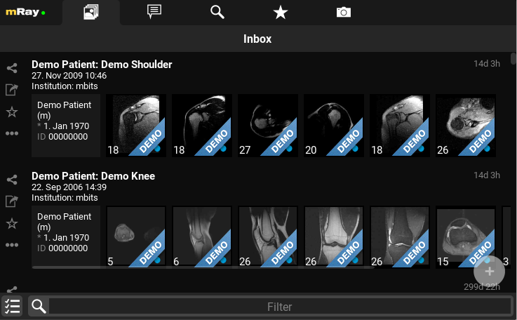
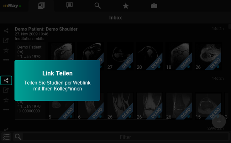
Sie können Ihre Bilddaten über das “Teilen” Symbol in der Bedienleiste links teilen.
Dafür kann es mehrere Ursachen geben:
- Ihr Browser ist nicht für die Darstellung geeignet
Für die Darstellung geeignete Browser:
Google Chrome, Firefox, Microsoft Edge, Safari - Fehlende Verbindung zum Internet
Stellen Sie sicher, dass Sie mit dem Internet verbunden sind - Anmeldedaten wurden falsch eigegeben
Folgen Sie den Anweisungen auf Ihrem Ausdruck, dort finden Sie Ihren Zugangscode und Ihre PIN.
Sie haben jederzeit die Möglichkeit, auch ohne den QR Code die Bilddaten abzurufen. Folgen Sie hierzu den Anweisungen auf dem Ausdruck. Dort finden Sie eine Internet-Adresse, die Sie auffordert Ihren Zugangscode und PIN einzugeben.
Wenn Ihre Bilddaten nicht mehr angezeigt werden, ist deren Gültigkeit abgelaufen. Damit Sie wieder Zugriff auf Ihre Bilddaten erhalten, fragen Sie bitte bei Ihrer Praxis nach einer Verlängerung Ihrer Bilddaten nach.
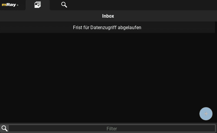
Wenn Ihre Bilddaten nicht mehr angezeigt werden, ist deren Gültigkeit abgelaufen. Damit Sie wieder Zugriff auf Ihre Bilddaten erhalten, fragen Sie bei Ihrer Praxis nach einer Verlängerung Ihrer Bilddaten nach.
Zwischen der Bildgebung und einem vollständigen Befund besteht immer ein zeitlicher Versatz. Der Befund erscheint, sobald er freigegeben wurde. Versuchen Sie es etwas später erneut, oder wenden Sie sich an Ihre Praxis, sollte er weiterhin fehlen.
Öffnen Sie Ihren Befund und gehen Sie rechts unten auf folgendes Symbol ![]() , um diesen herunterzuladen. Sie können das PDF anschließend wie gewohnt ausdrucken.
, um diesen herunterzuladen. Sie können das PDF anschließend wie gewohnt ausdrucken.
mRay FAQ - Datensicherheit
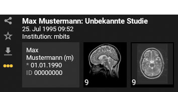
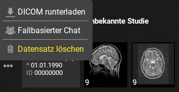
Um Ihre Daten in mRay zu löschen, klicken Sie einfach auf die drei Punkte neben dem Datensatz und wählen Sie “Datensatz löschen” aus.
Um Ihren mRay Privat-Account zu löschen, klicken Sie auf das mRay Logo oben links. Anschließend klicken Sie auf die Zahnräder neben Ihrem Namen, um zu Ihren Profileinstellungen zu gelangen. Unter “Account-Management” finden Sie die Option, Ihren Account zu löschen.
